Previous Topic

Next Topic

Book Contents

Book Index

Marker Guidance

The automatic myocardium detection can be difficult, especially if the image quality is poor, if there are area of reduced uptake, or if there is no clear separation between myocardium and liver. To make the automatic outlining more robust, the user can specify anatomical landmarks. The markers should be placed at the basal points and the apex of the left ventricle as illustrated below.

PCARD Markers for AutoVOI

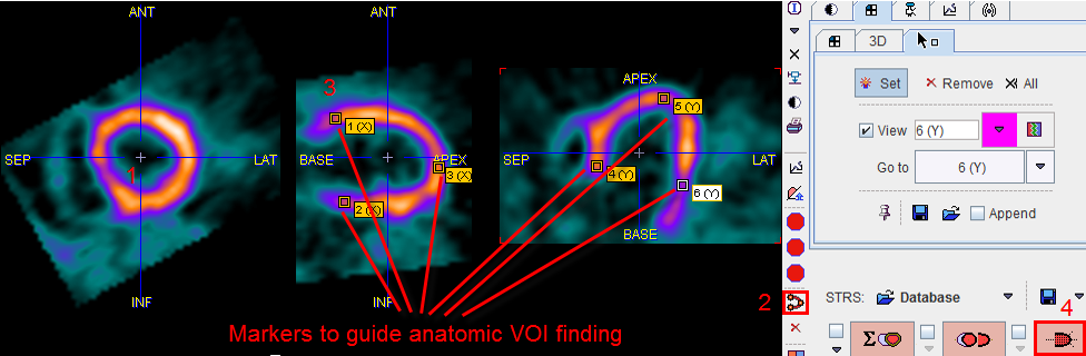
Marker Definition Procedure

  1. Navigate the images by clicking into the LV centre until the LV extremes are visible.
  2. Select the PCARD LV Markers button. The tabs in the image controls area are switched to the Markers tab.
  3. Click at 5 to 6 points in the long axis views to set markers as indicated in the illustration above. Note that to avoid setting markers when clicking into the image the Set toggle button in the Markers tab must be switched to inactive.
  4. Finally select the contouring button Button Myocardium Contouring to restart the automatic contour detection algorithm.
  5. If the outlining result is not satisfactory, just select new marker points and repeat, or enter the procedure for manually editing of the VOIs.

Note: The image information above the basal marker points will completely be disregarded in the outlining procedure. The apex point, however, will be adjusted based on the image content. This marker-guided procedure can be applied even after VOIs have been defined.