![]()
![]()
![]()
![]()
|
|
|
|
|
The TAC of the lungs provides a reasonable approximation of the shape of the blood curve. It is obtained in a lung VOI which has traditionally been outlined in matched transmission images because of poor anatomical information in the early water images. With images acquired using current scanners the lung VOIs may directly be outlined in the PET images.
Please perform the following steps for an analysis using CT images.
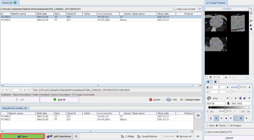
 VOI button. In the appearing VOI construction dialog make sure to switch to the transmission images and draw regions in the left and right lung.
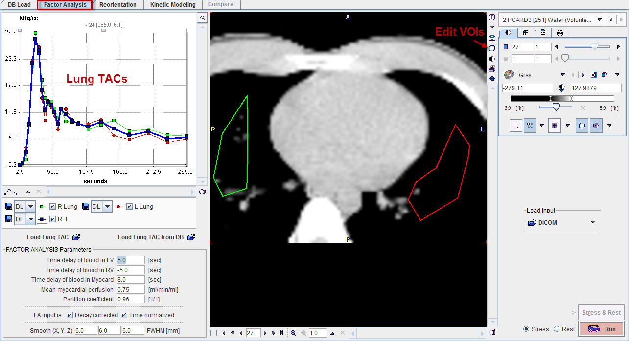
Notes:
1. It is important that the acquisition times of the dynamic images are correct.
2. If the acquisition has been performed using a hybrid PET/CT scanner it should be possible to use the CT images for defining the Lung VOIs, because PMOD relates the VOI to the scan landmark which is normally identical in both scans.
If the CT images are not required for the location of the lungs, VOI outlining can be directly performed in the water images. In this case it is even possible to load Stress and Rest images, outline the VOIs, and activate the Stress & Rest button.
If the lung TAC is already available as a file the loading of the transmission data and the VOI outlining is not necessary. In this case it is sufficient to load the dynamic H215O-PET images, then load the lung TAC with one of the buttons Load Lung TAC or Load Lung TAC from DB, Then the FA can immediately be started.