![]()
![]()
![]()
![]()
|
|
|
|
|
The rBV ( Autorad) model is intended for the quantitative assessment of the regional blood volume (rBV). The required measurements are a static C15O PET scan and external blood sampling. After a bolus inhalation of C15O inhalation an equilibration period should be allowed for 5 min. Then, blood data are sampled while a static PET acquisition is performed. The rBV can finally be calculated by dividing the PET activity by the integrated blood activity.
Acquisition and Data Requirements
Image Data |
A static PET data set representing the equilibrium brain activity during C15O inhalation. If more than one frame has been loaded, the average PET activity is calculated during the pixel-wise calculations. |
Blood Data |
Blood activity sampled at a peripheral artery after equilibration from beginning until the end of the PET acquisition. |
Blood Preprocessing
The only available blood preprocessing option is decay correction.
Model Preprocessing
Just reads numerical data needed for pixel-wise processing in two input fields.
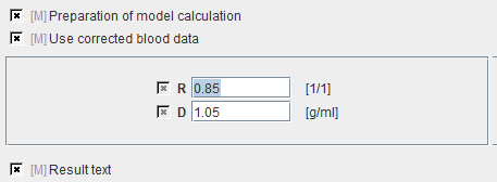
R |
Haematokrit Ratio Small/Large Vessels. Mean ratio: 0.85. |
D |
Density of brain tissue: 1.05 [g/ml]. |
Map Parameters

rBV |
Regional blood volume in [ml/100g]. It is calculated as: |
Reference
1. Mintun MA, Raichle ME, Martin WR, Herscovitch P: Brain oxygen utilization measured with O-15 radiotracers and positron emission tomography. J Nucl Med 1984, 25(2):177-187.
Note: The main focus of the cited reference is the calculation of regional brain oxygen extraction (CMRO2). That model requires the pixel-wise knowledge of the rBV and the rCBF. These functional maps could be determined with models supported in this software. The calculation of the CMRO2, however, is currently not available.