![]()
![]()
![]()
![]()
|
|
|
|
|
VOI templates are usually image files which represent a standard anatomy. The values in the image pixels are label numbers, and each label number corresponds to a certain anatomical region. For example, the labels in the AAL template [1] are described by a list in the form
FAG Precentral_L 2001FAD Precentral_R 2002F1G Frontal_Sup_L 2101F1D Frontal_Sup_R 2102F1OG Frontal_Sup_Orb_L 2111F1OD Frontal_Sup_Orb_R 2112F2G Frontal_Mid_L 2201F2D Frontal_Mid_R 2202F2OG Frontal_Mid_Orb_L 2211F2OD Frontal_Mid_Orb_R 2212etc.
and the template images showing the label regions look as illustrated below (interpolation disabled).
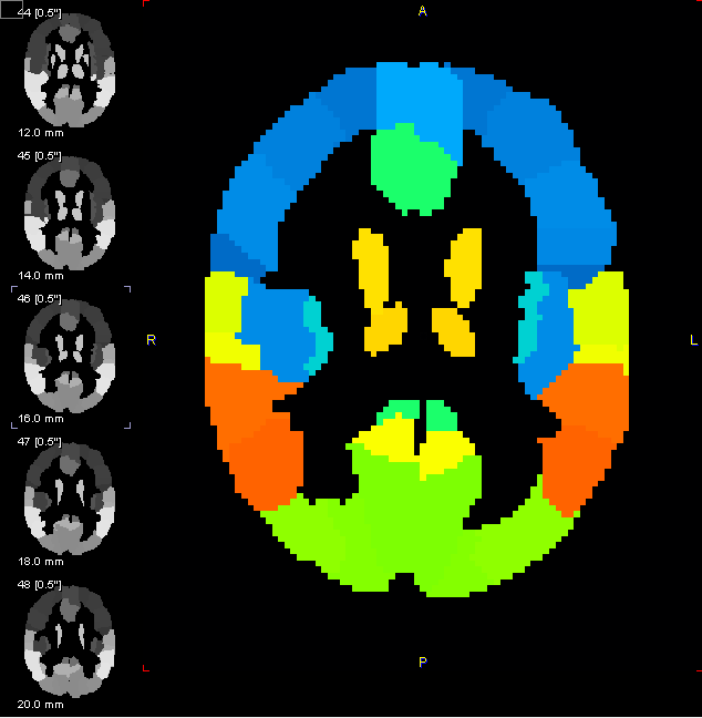
The anatomy of the AAL template corresponds to the spatially normalized, single subject, high resolution T1 MRI data set provided by the Montreal Neurological Institute (MNI) (Collins et al., 1998) in HFS orientation (radiological convention). All images which have been spatially normalized to a HFS template in the PMOD image fusion tool can be used together with the AAL template.
Notes: There is a slight asymmetry in the AAL template VOIs which corresponds to the natural asymmetry of normal brains and which is also part of the MNI template. The resolution of the AAL atlas is 2mm in all directions. Stereotactic data with other resolution must be interpolated to 2mm and brought into HFS orientation to apply the AAL template within PMOD.
[1] Tzourio-Mazoyer N, Landeau B, Papathanassiou D, Crivello F, Etard O, Delcroix N, et al. Automated anatomical labeling of activations in spm using a macroscopic anatomical parcellation of the MNI MRI single subject brain. Neuroimage 2002; 15: 273-289.
[2] Collins DL, Zijdenbos AP, Kollokian V, Sled JG, Kabani NJ, Holmes CJ, Evans AC. Design and construction of a realistic digital brain phantom. IEEE Trans Med Imaging. 1998 Jun;17(3):463-8.