![]()
![]()
![]()
![]()
|
|
|
|
|
This auxiliary model is aimed at receptor tracers for which a reference tissue without specific binding is available. It allows getting a quick estimate of the binding potential BP using two different methods as described by Ito et al. [24].
At the time, when specific binding C2 peaks, its derivative equals zero (transient equilibrium) and BP=k3/k4=C2/C1 (non-specific/specific binding). In principle, however, neither C1 nor C2 of a tissue TAC are known. It is assumed that specific binding is equal among tissues, so the TACns from a tissue without specific binding (reference) is regarded as C1, and C2 is calculated by TAC- TACns.
Two different ratios are calculated by the Tissue Ratio Methods model:
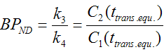
This ratio is calculated for all times, but it is only valid at the time of transient equilibrium. There is no automatic routine for detecting this peak at the moment. However, the user can easily find this time by looking at the Tac - Reference curve (enable check box).
The second ratio calculated is called the interval method in [24].
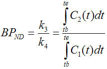
Here, the two curves are integrated in a time interval which must include the time of transient equilibrium (tb=9min. te=45 min for raclopride, where the time of transient equilibrium is in the range 20 to 24 min [24]).
Implementation Notes
Abstract [24]
"Several approaches have been applied for quantification of D2 dopamine receptors in positron emission tomography studies using [11C]raclopride. Initial approaches were based on analyses of data obtained after rapid bolus injection of [11C]raclopride. A continuous infusion paradigm has more recently been applied. The current study compares these approaches in healthy men. Two positron emission tomography measurements were performed in each of six healthy men, the first with rapid bolus injection and the second with continuous infusion of [11C]raclopride. In rapid bolus injection, the binding potential was calculated by the following methods. One approach is the kinetic analysis using the standard three- compartment model. Another is to define a transient equilibrium at the moment when the specific binding reaches its maximum. In continuous infusion, binding potential was calculated by using time-activity data at equilibrium condition. All methods gave almost identical binding potential, representing cross-validation of these methods. The continuous infusion method can provide "true" equilibrium condition. The kinetic analysis is a sophisticated approach but requires determination of an arterial input function. The transient equilibrium method thus is suitable for routine clinical research, since it does not require determination of an arterial input function."