![]()
![]()
![]()
![]()
|
|
|
|
|
SEGMENTS Image
The hierarchy of segments is organized as a "dynamic" series with increasing number of segments. The first "frame" has only a single segment which corresponds to the whole body mask. The second segment decomposes the area of the body mask into a background and a single segment, the third shows two segments, and so on, as illustrated below.
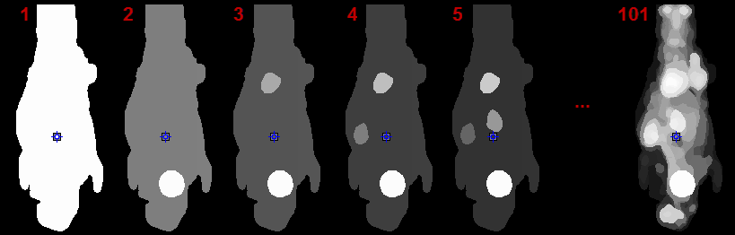
The number of segments can be adjusted by entering the number into indicated fields or dragging the slider.
Note: On each level, the segments decompose the body mask into disjoint, compact volumes.
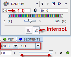
Per default the RANDOM color table is applied for showing the segments decomposition with the aim of separating neighboring segments. Interpolation is switched off to avoid distracting color effects at segment boundaries.
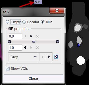
The MIP representation of the SEGMENTS image is a valuable tool for detecting segments of interest. To have it available please make sure the  tab is selected. Furthermore, the full range should be enabled for the MIP.
Reference Image
The reference image which is used as a backdrop for the segments can be selected as illustrated below. The dynamic PET as well as the averaged PET (AVR), are always available, and the anatomical image optionally.
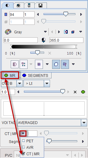
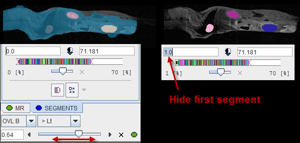
Image Fusion
Per default the fusion mode is set to OVL B >Lt, meaning that only the segments above the lower threshold are shown in the image. It may be helpful to set the threshold to 1.0 in order to hide the first "background segment". Please use the blending slider to inspect the reference image below the segments.
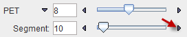
Navigational Elements
The number of segments is best changed in the lower right slider area. The recommended procedure is to set the Segment number to 1, and then gradually increase it by the increment arrow.
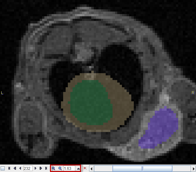
The easiest way to localize segments in the image is via the MIP image: Clicking at a segment will update the orthogonal image planes with intersection at the segment. To center the intersection point on the segment please click on the slice images, or use the mouse wheel for scrolling slices.
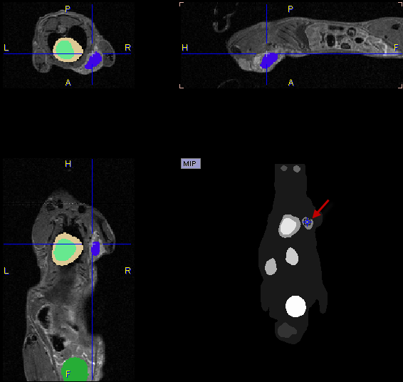
Close examination might require switching the image layout from the orthogonal layout to a single plane layout and some zooming, and then adjusting the blending. There are zooming elements available below the image, but the image can also be zoomed by the mouse wheel when holding down the Ctrl key.